Digital Pelvic Phantom
|
This repository contains DICOM images derived from MRI and CT scans of a single patient. The
patient had three fiducials implanted in the prostate and was imaged and treated with a full
bladder on a Tomotherapy unit. The images in the series were derived from the CT taken for
treatment planning
(Pelvis0)
and six of the daily Megavoltage CT’s taken immediately prior to
treatment
(Pelvis1 through Pelvis6)
. The prostate and urethra contoured on the MRI, bone
contoured on the planning CT, and bladder, rectum, bowel, bowel gas, and soft tissue contoured
in the CT images, were each assigned a single Hounsfield Unit: -1000 for air and bowel gas, 35
for prostate, 36 for soft tissue, 37 for small bowel, 38 for rectum, 39 for small bowel, 52 for
bladder and urethra, and 1296 for bone. The bone contours from the planning CT were
transferred to the daily CT’s following rigid registration. The position of the most inferior and
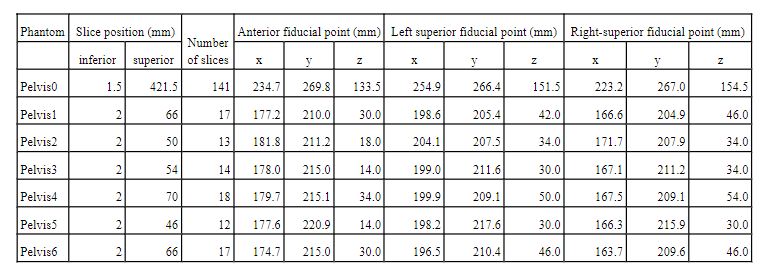
most superior axial slices, number of slices, and the position of the fiducials in each phantom
(x is right-left, y anterior-posterior, z superior-inferior)
is shown below
(Table I of the reference): |
 |
 |
|
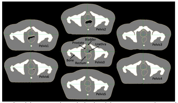
Figure: Digital phantom series showing the slice through the prostate nearest to the centroid of
the fiducials for all seven phantoms. From Faddegon et al, 2023. |
| Reference: Bruce Faddegon, Martina Descovich, Katherine Chen, Jose Ramos Mendez, Mack Roach III, Niklas Wahl, Paige Taylor, Keith Griffin, Choonsik Lee, “A Digital Male Pelvis Phantom Series Exhibiting Day-To-Day Anatomical Variation,” Medical Physics, Dec 2023. https://doi.org/10.1002/mp.16865 |